


A(nother) visual tour of the lumbar nerve roots!

Thanks for reading the 51st edition of my newsletter. This newsletter tracks my research as I write a book about sciatica, and another one (out very soon) about cauda equina syndrome.
By the way, if you’re not really enjoying these emails, you should unsubscribe! I hate the idea that they’re just clogging up someone’s inbox. Just scroll to the bottom and click ‘unsubscribe’ 👍
Hi everyone,
We were hoping to make the CES book available to buy this week, but there are still one or two technical things we need to clear up. It's a shame, but it shouldn't take too long and we hope it'll be out in the next couple of weeks...
Here’s a sneak peak though:

In the meantime, I thought I would re-write an old post, 'a visual tour of the lumbar nerve roots', adding some more explanations and some lovely spinal pictures I've found since I first wrote it over two years ago...
Here we go!
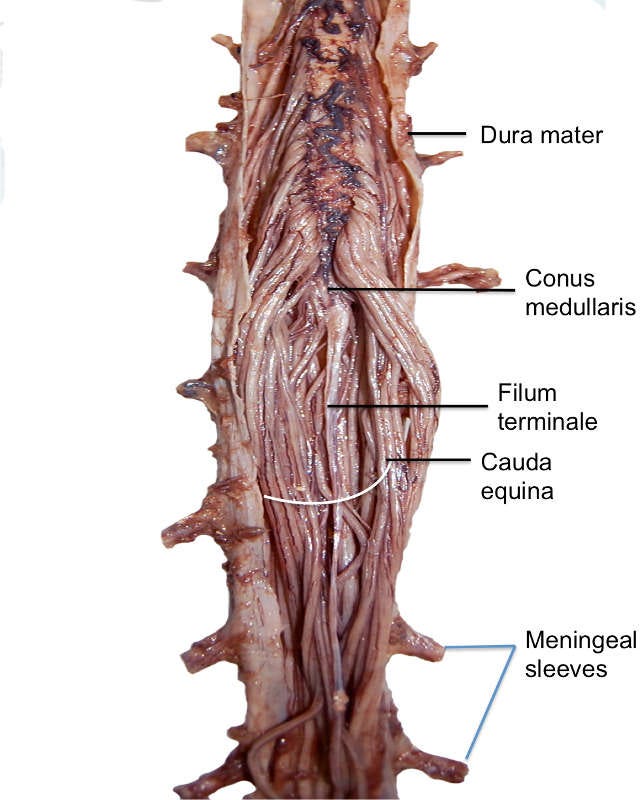
Let’s start at the top. The spinal cord ends at about the L1 vertebral level (higher than most people think!). The cord tapers to terminate at the conus medullaris:

As you can see below, the nerve roots first bud off from the spinal cord not as fully formed roots, but rootlets:

After the rootlets bud off from the spinal cord, they bundle up to become the nerve roots that make up the hanging tail of the cauda equina. Dorsal rootlets stick together and ventral rootlets stick together, making separate dorsal sensory roots and ventral motor roots.
At the lumbar spine, the spinal cord is small, about the size of a little finger. And the roots are smaller still, between two and four millimeters thick.
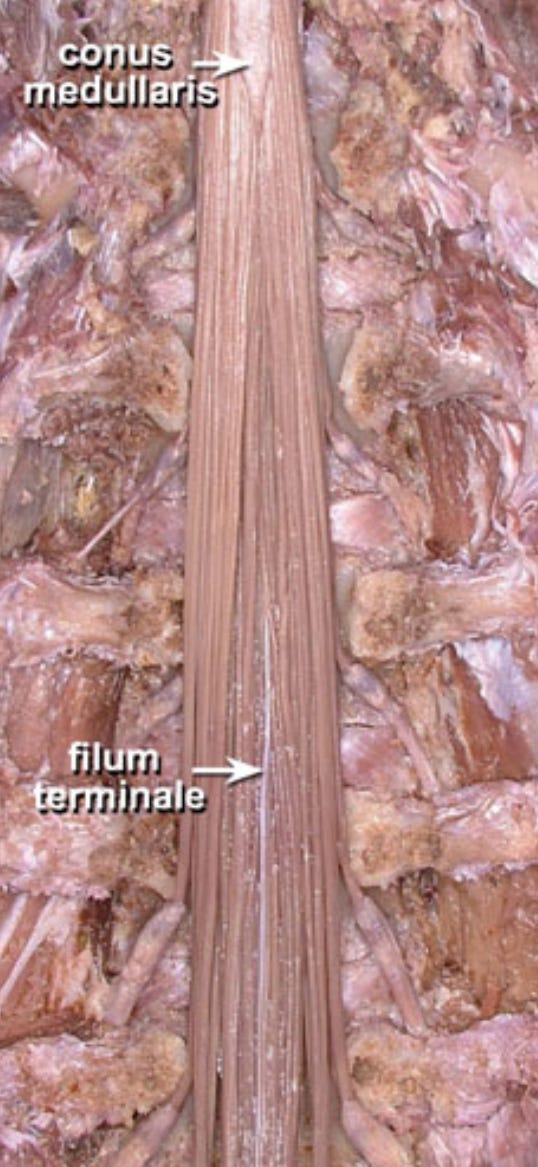
When an anatomist is laying the roots out or holding them they seem (to me…) like tangled spaghetti, hanging off a fork…
In the body, they are guided and held by ligaments and connective tissue, so they have a more orderly and linear appearance... (like uncooked spaghetti still in the packet! Although of course they are soft and pliable, with the consistency of rubber)

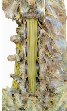
Everything we have seen so far is taking place inside the protective thecal sac. The thecal sac is made up of the same meninges that cover the brain and spinal cord. Here’s a similar view to those above, but with the thecal sac left on:
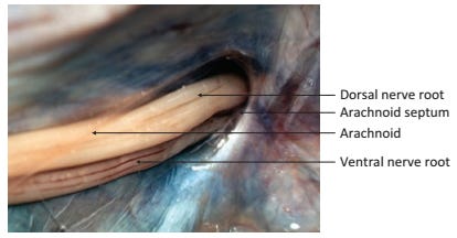
And here's the thecal sac in cross section:

And another cross section view:

Okay, so the nerve roots travel down through the thecal sac as the cauda equina. Next, let’s look at how, at each spinal level, one sensory and one motor root per side pair up and branch off together. They are on their way out of the spinal column to go forth and innervate their particular patches of the low back and leg.

As they branch off, the pair of roots take with them a portion of the thecal sac, which will now form what's called the nerve root sleeve:

In the short part of their course that you can see in the picture above, after they leave the thecal sac and before they leave the spinal column completely, the nerve roots are at their most vulnerable. This is partly because they don't have the space to maneuver that they had in the thecal sac, because the nerve root sleeve binds them quite tightly. Additionally, there are ligaments here that stop them from moving much:

This fixity makes them vulnerable to anything that might compress, stretch, pin or twist them—like a disc herniation. Not only that, but the root sleeve is less tough than the layers of connective tissue that protect the peripheral nerves proper when they get out into the lower limb…
It’s also at this point that the nerve roots pass through the intervertebral foramen. And it’s here in the foramen that the dorsal root ganglion usually resides. The lumbar ganglion is about the size of the fingernail on your little finger.

Inside the dorsal root ganglion are the cell bodies of the sensory nerves. These cell bodies manufacture everything that their sensory nerve needs to function. (See my previous post for more on the enigmatic ganglion!)
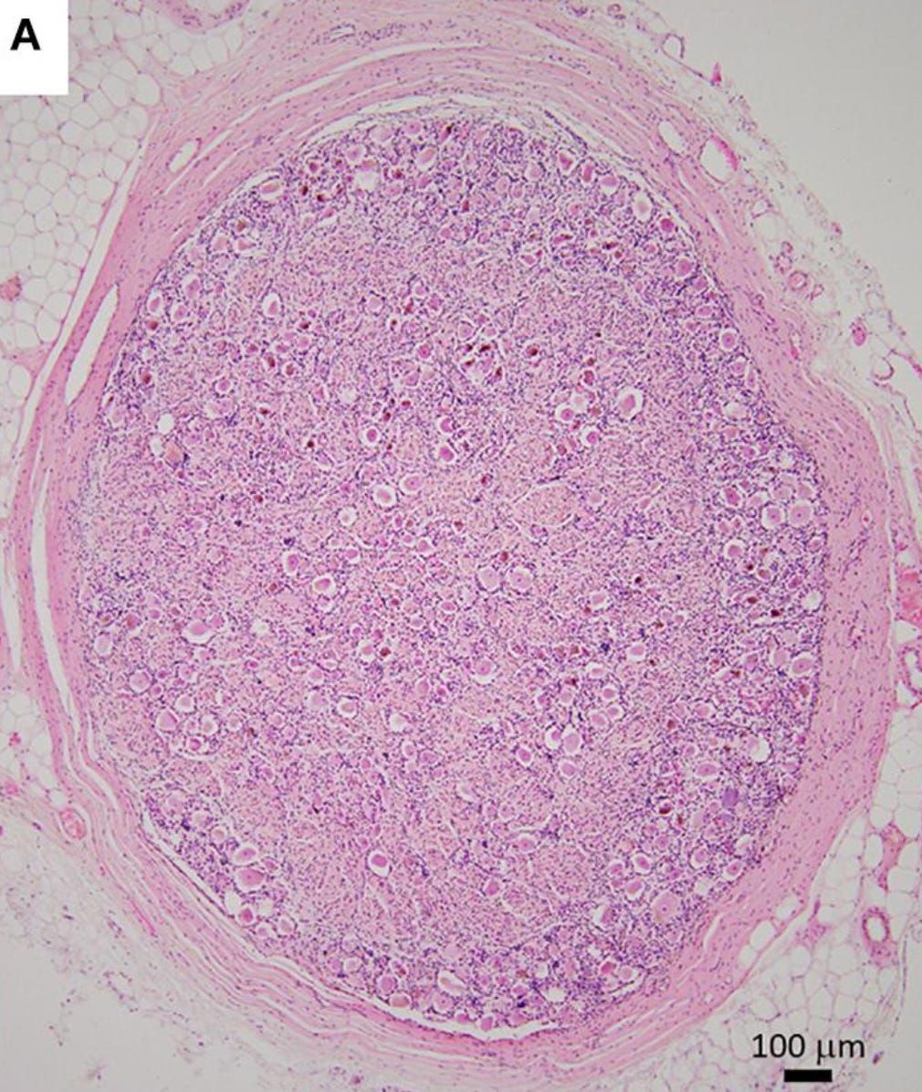
(This is a good place to remember that the dorsal root ganglion is just one tiny blip on a bloody gigantic sensory neuron)

Although the ganglion causes the root to thicken, the root still does not take up more than about a third of the space in the foramen. The rest of that space is taken up by blood vessels, ligaments, the sinuvertebral nerve and cushioning fat, as you can see in this side view:
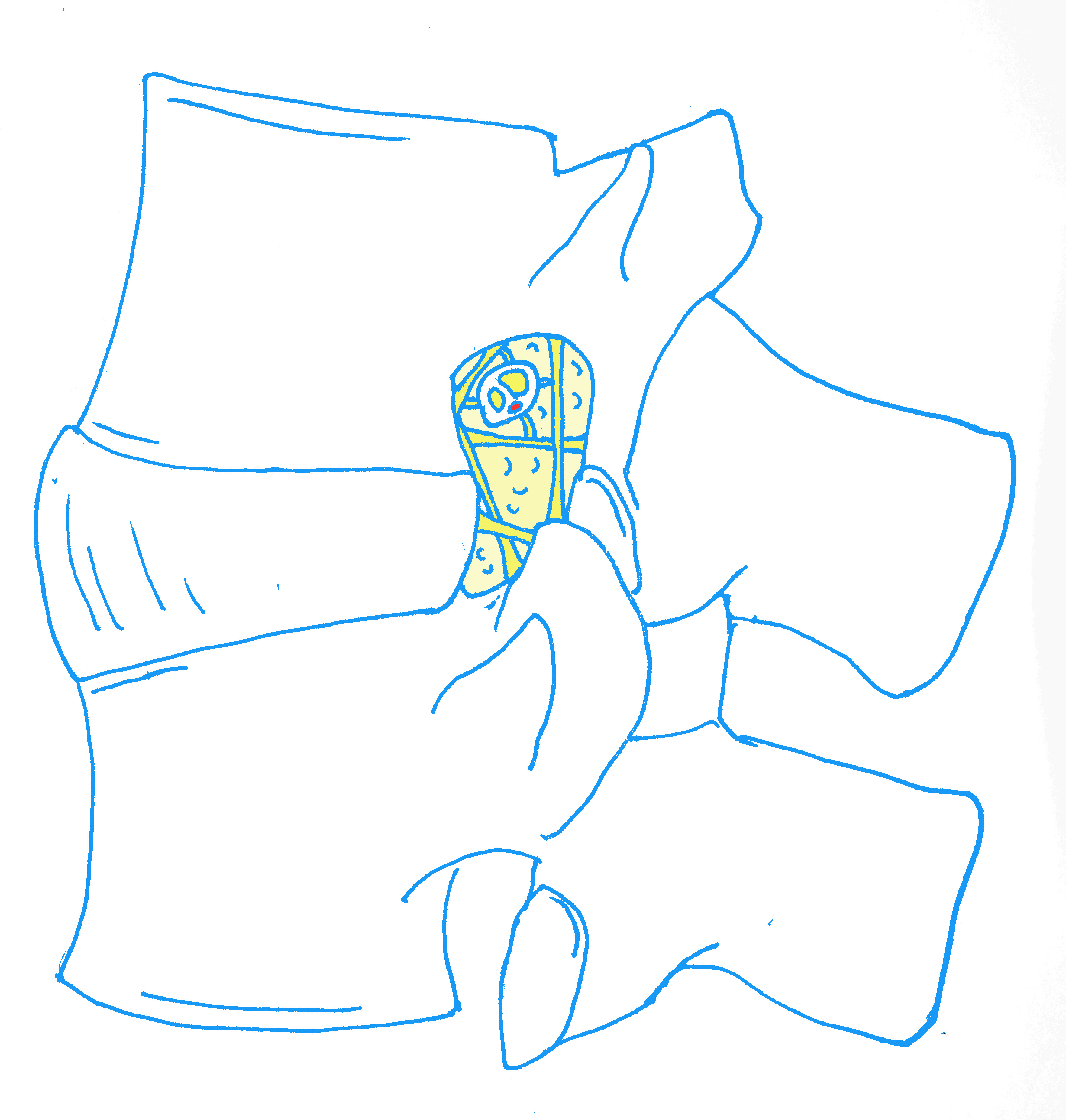
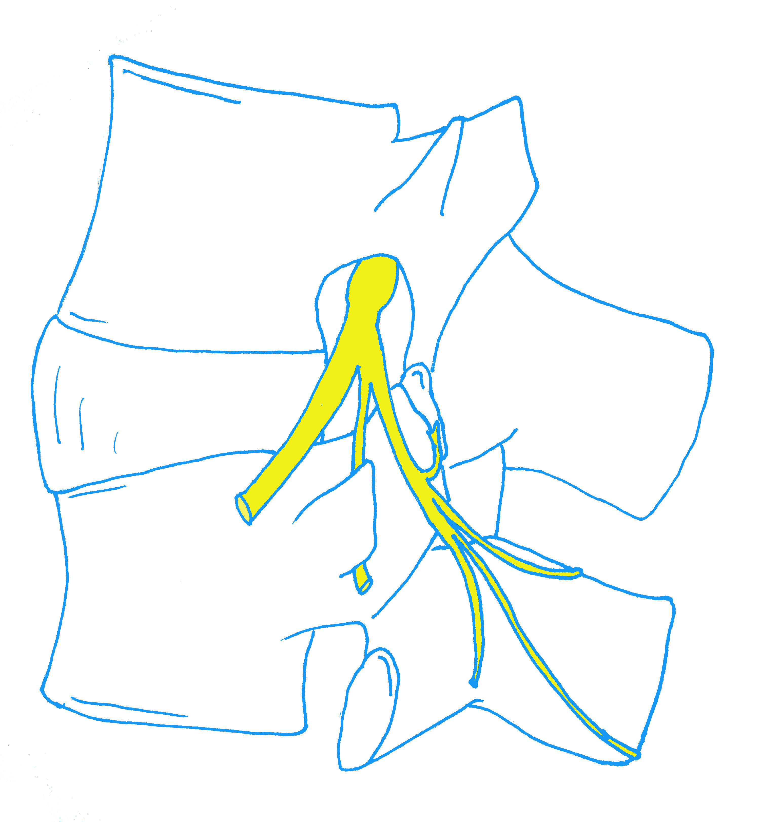
As a bit of a summary pic, here’s an illustration from my book of the sensory and motor nerve roots, with the DRG, inside the root sheath, passing through the intervertebral foramen.

As you can also see in that picture that, after the dorsal root ganglion, the pair of nerve roots undergo a major change: they are finally woven together into the mixed spinal nerve. This spinal nerve is remarkably short. Immediately outside the foramen, it branches off into the two rami, which will serve the structures of the spine and the lower limb. We have now left the nerve roots behind!
Now let’s look at the blood supply, which is a very under-rated factor in radicular syndromes. The descending aorta sits on the front of the vertebrae and at each spinal level, a pair of arteries branch off and course backwards around each side of the vertebrae.

As they make their way backwards, they send off a branch into the intervertebral foramen. This is the distal radicular artery. As it enters the foramen, the distal radicular artery pierces the spinal nerve and courses up, splitting to enter both nerve roots, forming a plexus around the ganglion and continuing proximally towards the spinal cord. It is through the distal radicular artery that the heart pumps blood up the nerve roots, towards the spinal cord. (There is also a proximal radicular artery coming back down in the other direction).

Blood is drained from the roots by radicular veins. Inside the nerve root, the veins run in spirals. Outside, they form large, thick plexuses around the roots.

It's easy to see how adding pressure to this vascular system can prevent blood from pumping in and draining out properly, starving the roots of oxygen.
Lastly, a quick word on disc herniations (maybe worth another visual tour email in themselves?)…
If you don’t already know the answer, here’s an exercise: look at this picture again and ask yourself which nerve root is most likely to be injured if there is an L5/S1 herniation:
The answer is the S1 nerve root, which could be contacted just as it’s leaving the thecal sac. And for an L4/5 disc herniation, the L5 nerve root will be affected in the same position, just as it’s leaving. This is a common point of confusion: disc herniations tend to affect the nerve root of the level below, as it’s ‘transiting’ past the disc. The nerve root at the level of the herniation is generally out of danger, as it’s already ‘exiting’ some way from the disc (except in the case of far lateral herniations).
As a final picture, here’s a fantastic view where you can see pretty much everything we’ve just looked at. The photo is from Jose Paulo Andrade, on twitter.
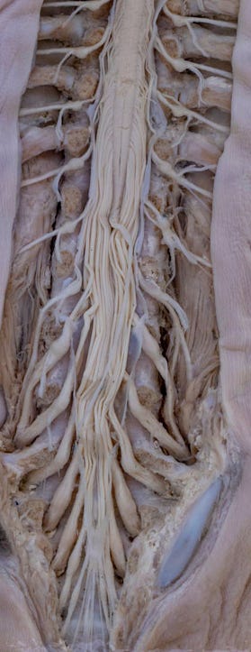
Oh, and one last picture where you can really see why it’s all named after a horse’s tail:

I hope that was an interesting diversion. Certainly much better than the version from two years ago, I think.
If you did find this interesting, please do share it with someone else who might.
Let’s see if we’re in a position to release the CES book next week… I hope so! 🤞
Til next time,
Tom